Journal of JCIC

Online edition: ISSN 2432–2342
JCIC学会事務局 JCIC学会事務局
〒162-0801東京都新宿区山吹町358-5アカデミーセンター Academy Center, 358-5 Yamabuki-cho, Shinju-ku, Tokyo 162-0801, Japan
Journal of JCIC 5(2): 19-23 (2021)
doi:10.20599/jjcic.5.19

原著原著

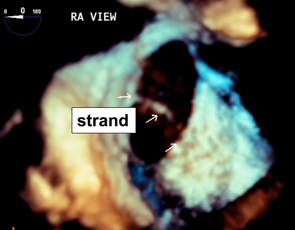
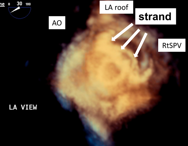
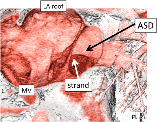
紐状構造物(strand)を支持力の一部として利用した経皮的心房中隔欠損閉鎖術Percutaneous Transcatheter Closure of Atrial Septal Defect Using Strand as a Part of the Supporting Margin

静岡県立こども病院 循環器科Department of Cardiology, Shizuoka Children’s Hospital

受付日:2020年5月27日Received: May 27, 2020
受理日:2021年1月29日Accepted: January 29, 2021
発行日:2021年3月25日Published: March 25, 2021
HTMLPDFEPUB3

【背景】多孔性心房中隔欠損(multi-fenestrated atrial septal defect; MFASD)の類縁病変として1つの欠損孔が紐状構造物(strand)により,まれに2分割されることがあり,経皮的心房中隔欠損(atrial septal defect; ASD)閉鎖術の際に手技上の特別な配慮が必要となる.閉鎖術の際にstrandを支持力のある辺縁の一部として利用できる可能性とその効果について検討した.

【方法】当院で経験したASD症例のうち,経食道心エコー法でstrandを認めた8例を後方視的に検討した.最大幅3 mm以下の構造物をstrandと定義した.

【結果】strandは斜走する形で存在し,主欠損孔は5例で前上方に位置.5例でカテーテル治療,3例で外科手術を選択した.経食道心エコーをガイドとして,主欠損孔にて2段階のバルーンサイジング(短絡が僅かに残る程度の弱バルーンサイジング法,及びwaistができる程度の強バルーンサイジング法)を行い,strandを含めた辺縁の伸展性を評価.強バルーンサイジング法を指標として閉鎖栓を留置した.1例のみ治療6か月後に残存短絡を極少量認めた.

【結論】strandが辺縁の一部として閉鎖術に利用できる可能性があり,存在する位置と合併する辺縁欠損及び辺縁全体の伸展性を評価することが重要と考える.

【Background】In multi-fenestrated atrial septal defects (MFASD), one defect may rarely be divided into two due to a strand. Therefore, we should be careful during percutaneous transcatheter closure of atrial septal defects (ASD). We investigated the possibility and effects of using a strand as part of the supporting margin during a catheter intervention.

【Methods】This retrospective study included eight cases of ASD with a strand that were treated at our hospital. A structure with a maximum width of 3 mm or less was defined as a strand.

【Results】In all patients, the strands ran diagonally, and five patients had the main defect located in the antero-superior part. Five patients underwent transcatheter device closure and three patients underwent surgical closure. Under transesophageal echocardiography monitoring, we performed two-stage balloon sizing using the weak balloon sizing method where a slight shunt flow remains and the strong balloon sizing method which involves complete disappearance of the shunt flow and a slight balloon waist in the main defect. We then evaluated the compliance of the rim, including the strand. The closure devices were placed with reference to the strong balloon sizing method. Only one patient exhibited a small amount of residual shunt at the six months follow-up.

【Conclusion】The strand may act as a part of the margin for device closure. The defect position and deficient rim should be evaluated, in addition to the compliance of the supporting strand.

Key words: Atrial septal defect; multi-fenestrated; strand; Amplatzer Septal Occluder; Occulutech Figulla Septal Occluder

背景

複数個の欠損孔をもつ多孔性心房中隔欠損(multi-fenestrated atrial septal defect; MFASD)に対する経皮的心房中隔欠損(atrial septal defect; ASD)閉鎖術では,残存短絡のリスクや複数閉鎖栓の留置を要する可能性があり,手技上の特別な配慮が必要である.一方,心房中隔欠損症で経食道心エコー検査(transesophageal echocardiography; TEE)を実施すると,まれに1つの欠損孔が紐状構造物(strand)によって2分割された所見を認めることがある(Figs. 1, 2).経皮的心房中隔欠損閉鎖術の際,このstrandを支持力のある辺縁の一部として利用できる可能性があるが,治療に際してその適応や留置手技の判断に苦慮する場合がある.

Journal of JCIC 5(2): 19-23 (2021)

Fig. 1 TEE*1) (3D): Atrial septal defect divided by strand

*1) TEE: transesophageal echocardiography.

Journal of JCIC 5(2): 19-23 (2021)

Fig. 2 TEE (3D): transcatheter closure of atrial septal defect using strand

目的

心房中隔欠損閉鎖術におけるstrandの影響に関して,TEEを中心とする画像診断とともに検討する.

対象・方法

2011年1月~2019年12月の間に当院でカテーテル治療・外科手術を行った2次孔型ASDは全256例あり(内訳はカテーテル治療139例,外科手術117例),うち18例がMFASDだった.最大幅3 mm以下の紐状構造物をstrandと定義し,その他のMFASDと区別した.より大きな面積を持つ欠損孔を主欠損孔,それ以外を副欠損孔と定義した.事前に行ったTEEでstrandを認めた8例を対象とした.

MFASDに対してTEEで2段階のバルーンサイジング(①弱バルーンサイジング法:主欠損孔の短絡が僅かに残る程度 ②強バルーンサイジング法:主欠損孔の短絡が消失し軽度のwaistができる程度)を行い,strandを含めた辺縁の伸展性もあわせて評価した(Fig. 3).サイジングバルーンを通過させる孔は,主欠損孔を選択し,副孔でのバルーンサイジングは行わなかった.さらにASD全体を1つの大きな欠損孔とした径も計測し,閉鎖栓サイズ選択の参考にした.また,強・弱バルーンサイジング法でのバルーンサイジング径の差が大きい(1.5 mm以上)と辺縁の伸展性が低く(軟らかい),差が小さい(1.5 mm未満)と伸展性が高い(硬い)という性状判断を行った.また8例中1例は,他院で撮像したmulti-detector computed tomography(以下,MDCT)検査を参考に欠損孔の位置関係を確認した(当院では原則CT検査は行っていない).これらの計測を用いて,最終的に閉鎖栓の決定に際しては,主欠損孔に対する強バルーンサイジング法を指標とし,主欠損孔へ閉鎖栓の留置をおこなった.診療録をもとに,8例の形態,バルーンサイジングの結果,治療方法を後方視的に検討した.

Journal of JCIC 5(2): 19-23 (2021)

Fig. 3 balloon sizing

(A: weak balloon sizing method, B: strong balloon sizing method).

結果

8例の年齢は中央値10歳(3~62歳),体重は中央値34.0 kg(14.4~56.5 kg),性別は男2人女6人であった.カテーテル治療群は5例(Figs. 4, 5)外科手術群は3例(Figs. 6, 7)であった.それぞれの群の計測値は表の通りであった.

Journal of JCIC 5(2): 19-23 (2021)

Fig. 4 ASD (RA view, Catheter intervention group)

①deficient posterior rim ②deficient aortic rim, floppy postero-inferior rim ③deficient aortic and posterior rims, floppy atrioventricular valve rim ④deficient postero-inferior, aortic and superior rims, the septum primum is malaligned toward the left atrial side. ⑤deficient aortic rim, floppy postero-inferior and coronary sinus rims. SVC, superior vena cava; RtSPV, right superior pulmonary vein; IVC, inferior vena cava; CS, coronary sinus; TV, tricuspid valve; AO, aorta; RA, right atrium; LA left atrium; MV, mitral valve

Journal of JCIC 5(2): 19-23 (2021)

Fig. 5 Case④ MDCT*2) (4D)

*2) multi-detector computed tomography.

Journal of JCIC 5(2): 19-23 (2021)

Fig. 6 ASD (RA view, Surgical operation group)

⑥deficient aortic and inferior rim ⑦deficient aortic, superior and atrio- ventricular valve rims ⑧deficient aortic, posterior, postero-inferior rims (patient’s family hope to have surgical closure). Abbreviations are the same as in Fig. 4.

Journal of JCIC 5(2): 19-23 (2021)

Fig. 7 Case⑥ RA view, intraoperative photograph

Table 1
ASD全体の最大長径(平均)カテーテル治療群21.7±6.1 mm (11.0~29.0 mm)
外科手術群19.3±4.9 mm (12.9~24.9 mm)
ASD主欠損孔最大長径(平均)カテーテル治療群14.6±3.3 mm (7.9~18.0 mm)
外科手術群10.8±2.9 mm (6.5~13.7 mm)
バルーンサイジング径(平均)弱バルーンサイジング法18.2±4.7 mm (9.7~24.0 mm)
強バルーンサイジング法19.7±4.9 mm (10.7~25.0 mm)

strandは斜走する形で,5例が後下方側,3例が前上方側に位置していた.また7例でaortic rimが欠損していた.症例④はMDCT検査所見で後下縁から左房上壁側に偏位していた.ASD主欠損孔は全8例のうち5例で前上方に認め,うち3例はカテーテル治療を選択した.カテーテル治療を選択した症例は,全8例のうち5例で,強バルーンサイジング法を指標として閉鎖栓のサイズを決定した.閉鎖栓の選択は,Amplatzer Septal Occluder(ASO; AGA medical Corporation, Golden Valley, Minnesota)3例,Figulla Septal Occluder(FSO; Occlutech GmbH, Jena, Germany)2例だった.家族が外科手術を希望した1例と,strandが後下方に著明に偏位してstrandの支持力を提供できないと予測した2例は,後日開心術の適応と判断した.

考察

2次孔型の心房中隔欠損症に関して,James E. Lockらは解剖学的に以下の4つに分類している1).① virtual absence of septum primum such that the ASD was the entire fossa ovalis. ② deficiency of septum primum. ③ fenestrated septum primum-multiple ASDs. ④ fenestrations in a deficient septum primum-multiple ASDs.このうち,複数個の欠損孔をもつ多孔性心房中隔欠損症(MFASD)は約10%の割合で発生すると言われている2).MFASDは③・④のようにfloppyな1次中隔内に複数の欠損孔を認めるものである一方で,稀に1次中隔から2次中隔ないし左房天井に接続する紐状構造物(strand)を認めることがある.これはカテーテル治療を行う際に治療の妨げとなる場合がある.ASD欠損孔同士の間隔が7 mm以上あれば,短絡を残さないために複数の閉鎖栓を用いることが勧められるが3),間隔がそれ未満の場合には一つの閉鎖栓を如何に留置できるかを考慮する必要がでてくる.事前に経胸壁心エコー法や経食道心エコー法などの画像検査を用いて,空間的位置関係の把握を行うことは,治療戦略を考える上で非常に重要となる.3Dエコー検査の有効性も報告されている4, 5).過去の文献で調べうる限り,strandを切除して閉鎖栓を留置した報告はあるが6),strandを支持力の一部として使用してカテーテル治療を行った報告はなかった.ASD周囲縁のうち,後下縁欠損はカテーテル治療上の危険因子と言われている7).当院での治療戦略をまとめると,カテーテル治療はstrandが前上方側に位置する場合には主欠損孔が中央~前上方に位置してaortic rimが欠損していても閉鎖栓留置可能と判断し,主欠損孔が後下方に位置している場合にはpostero-inferior rimが広範囲に欠損していなければ留置可能と判断している.またstrandが後下方側に位置する場合,主欠損孔の位置に関わらず,それを支持力をもつpostero-inferior rimの一部として利用する方針としており,主欠損孔をバルーンサイジングする際に,strandの伸展性と壁在のpostero-inferior rim(多くの場合,寄って一体化する傾向がある)とを併せて,全体として伸展性を評価している.一方で,strandが後下方側により一層偏位しており,支持力が提供できないと判断した場合には外科治療を選択している.そして外来でのTEEで事前に診断がついた症例は,最終的にご家族の希望も反映した治療選択となる.

本検討では,前述のとおりASD周囲縁が欠損した症例の中でも,strandが著明に後下方に偏位してstrandの支持力が提供できないと判断した症例は外科手術適応と判断した.一方で,ASD主欠損孔の位置が心房中隔中央~前上方にあり,かつstrandの位置・伸展性次第ではASD周囲縁が欠損した症例でもカテーテル治療の適応と成り得る.

結論

経皮的心房中隔欠損閉鎖術の際,strandが辺縁の一部として利用できる可能性があり,存在する位置と合併する辺縁欠損及び辺縁全体の伸展性を評価することが重要と考える.

利益相反

日本小児循環器学会の定める利益相反に関する開示項目はありません.

本内容は第31回日本Pediatric Intervention Cardiology学会学術集会にて2020年1月23日に発表した.

引用文献References

1) Lock J, Rome J, Davis R, et al: Transcatheter closure of atrial septal defects experimental studies. Circulation 1989; 79: 1091–1099

2) Butera G, Romagnoli E, Saliba Z, et al: Percutaneous closure of multiple defects of the atrial septum: Procedural results and long-term follow-up. Catheter Cardiovasc Interv 2010; 76: 121–128

3) Awad S, Garay F, Cao Q-L, et al: Multiple Amplazter Septal Occluder Devices for Multiple Atrial Communications: Immediate and Long-Term Follow-up Results. Catheter Cardiovasc Interv 2007; 70: 265–273

4) Silvestry F, Cohen M, Armsby L, et al: American Society of Echocardiography; Society for Cardiac Angiography and Interventions: Guidelines for the Echocardiographic Assessment of Atrial Septal Defect and Patent Foramen Ovale: From the American Society of Echocardiography and Society for Cardiac Angiography and Interventions. J Am Soc Echocardiogr 2015; 28: 910–958

5) Georgakis A, Radtke W, Lopez C, et al: Complex Atrial Septal Defect: Percutaneous Repair Guided by Three-Dimensional Echocardiography. Echocardiography 2010; 27: 590–593

6) Carano N, Hagler D, Agnetti A, et al: Device closure of fenestrated atrial septal defects: use of a single Amplatz atrial septal occluder after balloon atrial septostomy to create a single defect. Catheter Cardiovasc Interv 2001; 52: 203–207

7) Fraisse A, Assaidi A, Kammache I, et al: TRANSCATHETER CLOSURE OF SECUNDUM ATRIAL SEPTAL DEFECT WITH DEFICIENT RIMS OTHER THAN THE ANTERO SUPERIOR USING AMPLATZER DEVICES. J Am Coll Cardiol 2013; 61: 498

This page was created on 2021-02-09T17:34:18.095+09:00
This page was last modified on 2021-03-22T13:29:18.000+09:00


このサイトは(株)国際文献社によって運用されています。